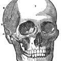
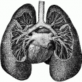
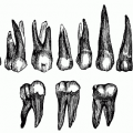
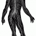
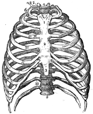
This is another great anatomical drawing from A Treatise on Anatomy, Physiology, and Hygiene. It was first published in 1852. This thorax drawing was included in the revised edition from 1858. It was written by Calvin Cutter, M.D.
This drawing depicts the thorax – the real name for what most of us refer to as the rib cage. It protects the lungs, heart and several large blood vessels like the aorta. At the back of the thorax is the twelve dorsal bones of the spinal column. Connecting to the dorsal bones are the ribs which are connected the sternum in the front. All of those are clearly labeled in this vintage anatomy drawing of the human thorax.
1, The first bone of the sternum, (breast-bone.) 2. The second bone of the sternum. 3, The cartilage of the sternum. 4, The first dorsal vertebra, (a bone of the spinal column.) 5, The last dorsal vertebra. 6, The first rib. 7, Its head. 8, Its neck. 9, Its tubercle. 10, The seventh, or last true rib. 11, The cartilage of the third rib. 12, The floating ribs.
This image is copyright free and in the public domain anywhere that extends copyrights 70 years after death or at least 120 years after publication when the original illustrator is unknown.
